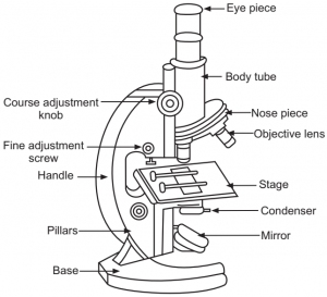
44 picture of compound microscope with labels
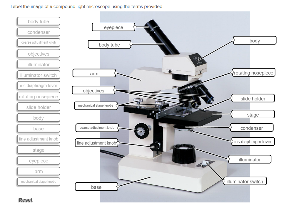
Microscope Types (with labeled diagrams) and Functions A compound microscope: Is used to view samples that are not visible to the naked eye Uses two types of lenses - Objective and ocular lenses Has a higher level of magnification - Typically up to 2000x Is used in hospitals and forensic labs by scientists, biologists and researchers to study micro organisms Compound microscope labeled diagram Label the microscope — Science Learning Hub All microscopes share features in common. In this interactive, you can label the different parts of a microscope. Use this with the Microscope parts activity to help students identify and label the main parts of a microscope and then describe their functions. Drag and drop the text labels onto the microscope diagram.
What is a Compound Microscope? - Study.com The body of the compound light microscope is the main part of the microscope, not to include the lights, focusing block, or stand of the microscope. The objective lenses and eyepiece are a part of ...

Picture of compound microscope with labels
› NATIONAL-GEOGRAPHIC-Dual-StudentNational Geographic Dual LED Student Microscope Aug 07, 2017 · Vanstarry Beginners Microscope Kit 40X-1000X for Kids & Students, Dual LED Lights and Cordless Capability, Illumination Lab Compound Monocular Microscopes with Optical Glass Lenses & 12 Slides 4.4 out of 5 stars 116 A Study of the Microscope and its Functions With a Labeled Diagram ... These labeled microscope diagrams and the functions of its various parts, attempt to simplify the microscope for you. However, as the saying goes, 'practice makes perfect', here is a blank compound microscope diagram and blank electron microscope diagram to label. Download the diagrams and practice labeling the different parts of these ... › 2022/10/12 › 23400986Microsoft takes the gloves off as it battles Sony for its ... Oct 12, 2022 · Microsoft pleaded for its deal on the day of the Phase 2 decision last month, but now the gloves are well and truly off. Microsoft describes the CMA’s concerns as “misplaced” and says that ...
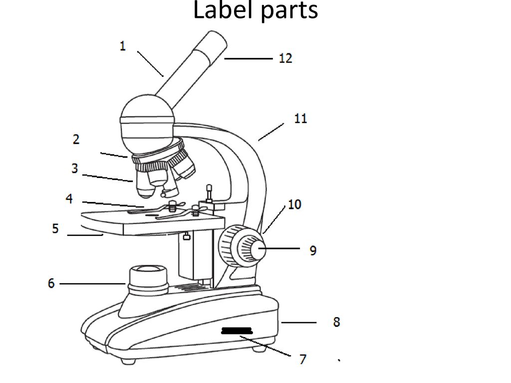
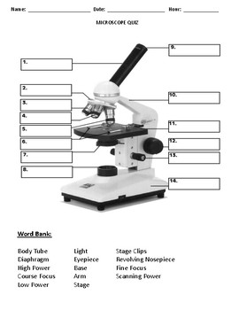
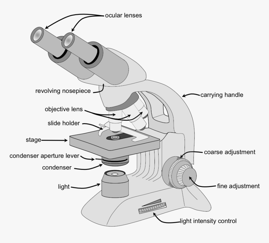
Picture of compound microscope with labels. Parts of the Microscope with Labeling (also Free Printouts) Picture Source: thorlabs.com 4. Objective lenses A microscope usually has 3 to 4 objective lenses with varying magnifications, usually 10 to 100x. Image 4: A closer look at the different objective lenses. Picture Source: microscopemaster.com 5. Knobs (fine and coarse) By adjusting the knob, you can adjust the focus of the microscope. Compound Microscope Parts The three basic, structural components of a compound microscope are the head, base and arm. Head/Body houses the optical parts in the upper part of the microscope. Base of the microscope supports the microscope and houses the illuminator. Arm connects to the base and supports the microscope head. It is also used to carry the microscope. Compound Microscope Parts - Labeled Diagram and their Functions Labeled diagram of a compound microscope Major structural parts of a compound microscope There are three major structural parts of a compound microscope. The head includes the upper part of the microscope, which houses the most critical optical components, and the eyepiece tube of the microscope. 962 Compound Microscope Stock Photos, Images & Pictures - Dreamstime Browse 962 professional compound microscope stock photos, images & pictures available royalty-free. Microscope in blue science medical technology laboratory background. Typical animal cell Center 400x. Microscopic micrograph of moth parts. Electronics. Electronics. Micrograph of lacewing insect.
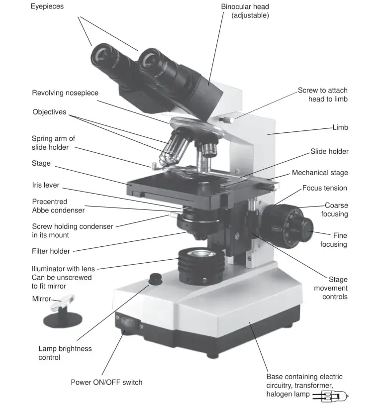
Compound microscope hi-res stock photography and images - Alamy RF2AMN35X-lens of compound microscope with medical sample slide in science laboratory white background RFS0C7CX-A common house fly leg under a compound microscope RF2B772F1-Method of illuminating compound microscope with gas lamp. Labels: C, Balloon flask filled with ammonium sulphate of copper and G, Welsbach mantle, vin Labelled Diagram of Compound Microscope The below mentioned article provides a labelled diagram of compound microscope. Part # 1. The Stand: The stand is made up of a heavy foot which carries a curved inclinable limb or arm bearing the body tube. The foot is generally horse shoe-shaped structure (Fig. 2) which rests on table top or any other surface on which the microscope in kept. Compound Microscope: Definition, Diagram, Parts, Uses, Working ... - BYJUS A microscope with a high resolution and uses two sets of lenses providing a 2-dimensional image of the sample. The term compound refers to the usage of more than one lens in the microscope. Also, the compound microscope is one of the types of optical microscopes. The other type of optical microscope is a simple microscope. Compound Microscope Parts, Functions, and Labeled Diagram Compound Microscope Definitions for Labels Eyepiece (ocular lens) with or without Pointer: The part that is looked through at the top of the compound microscope. Eyepieces typically have a magnification between 5x & 30x. Monocular or Binocular Head: Structural support that holds & connects the eyepieces to the objective lenses.
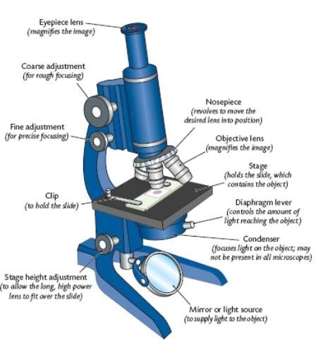
Parts of a microscope with functions and labeled diagram - Microbe Notes Microscope magnification measures the total enlargement of the image of an object. Magnification power is the product of eyepiece lens power and objective lens power. Q. Differentiate between the fine and the coarse adjustment knobs. Ans. The coarse adjustment knob moves the stage up and down to bring the specimen into focus. › solutions › lifeLive Cell Imaging | Solutions | Leica Microsystems Jun 01, 2022 · To perform successful live-cell imaging experiments, using the right platform is critical. When choosing an optical microscope for live‐cell imaging, the following 3 variables should be considered: detector sensitivity (signal‐to‐noise ratio), specimen viability, and image-acquisition speed. Compound microscope - their parts and function - Microscopy4kids Compound microscopes have more than one lens to generate high magnification images of flat, thin specimens. 2. Eyepiece (10x) and Objective lenses (4x, 10x, 40x, 100x) are two major optical parts of a microscope. 3. Total magnification power is calculated by multiplying the magnification of the eyepiece and objective lens. 4. Compound Microscope: Parts of Compound Microscope - BYJUS The parts of the compound microscope can be categorized into: Mechanical parts Optical parts (A) Mechanical Parts of a Compound Microscope 1. Foot or base It is a U-shaped structure and supports the entire weight of the compound microscope. 2. Pillar It is a vertical projection. This stands by resting on the base and supports the stage. 3. Arm
Below is a photo of a compound light microscope with labels ...
Compound Microscope Parts Made Easy - Microscope Detective A compound microscope employs a system of lenses and light to magnify the specimen. A light bulb illuminates the slide from below, and a series of lenses then magnify the sample. These are the objective lens near the slide and the eyepiece on top. The term "compound" refers to the compounding action of these two lenses.
Compound Microscope Review - ppt download
Compound Microscope Photos and Premium High Res Pictures - Getty Images Browse 992 compound microscope stock photos and images available, or search for light microscope or first compound microscope to find more great stock photos and pictures. of NEXT

The Microscope
Parts of a Compound Microscope and Their Functions - NotesHippo Compound microscope mechanical parts (Microscope Diagram: 2) include base or foot, pillar, arm, inclination joint, stage, clips, diaphragm, body tube, nose piece, coarse adjustment knob and fine adjustment knob. Base: It's the horseshoe-shaped base structure of microscope. All of the other components of the compound microscope are supported ...
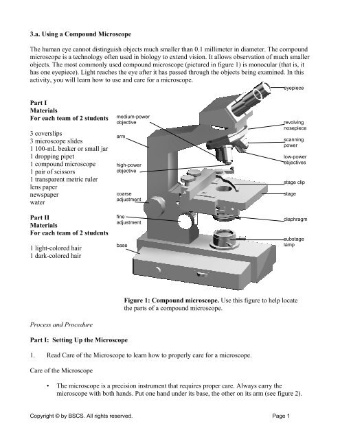
3.a. Using a Compound Microscope The human eye cannot ...
Compound Light Microscope Drawing - paintingvalley.com Are you looking for the best images of Compound Light Microscope Drawing? Here you are! We collected 36+ Compound Light Microscope Drawing paintings in our online museum of paintings - PaintingValley.com. ... Labeled Diagram Of C... 472x653 0 0. Like JPG. Light Microscopy Dra... 638x826 0 0. Like JPG. Microscope - Compoun... 236x305 0 0. Like ...
Microscope Parts & Specifications | Microscope World Resources
How to draw compound of Microscope easily - step by step I will show you " How to draw compound of microscope easily - step by step "Please watch carefully and try this okay.Thanks for watching.....#microscopedrawi...
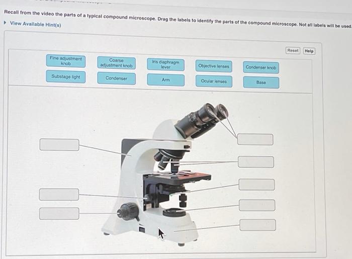
Solved Recall from the video the parts of a typical compound ...
› story › moneyUnbanked American households hit record low numbers in 2021 Oct 25, 2022 · The number of American households that were unbanked last year dropped to its lowest level since 2009, a dip due in part to people opening accounts to receive financial assistance during the ...

What is a Compound Microscope? | Microscope World Blog
Labeling the Parts of the Microscope | Microscope World Resources Labeling the Parts of the Microscope This activity has been designed for use in homes and schools. Each microscope layout (both blank and the version with answers) are available as PDF downloads. You can view a more in-depth review of each part of the microscope here. Download the Label the Parts of the Microscope PDF printable version here.

Parts of a microscope with functions and labeled diagram
Microscope Labeled Pictures, Images and Stock Photos photosynthesis. Diagram of the process of photosynthesis, showing the light reactions and the Calvin cycle. photosynthesis by absorbing water, light from the sun, and carbon dioxide from the atmosphere and converting it to sugars and oxygen. Light reactions occur in the thylakoid. Calvin Cycle occurs in the stoma. Neutrophil vector illustration.
Label Parts Of A Compound Microscope Teaching Resources | TPT
Parts of a Compound Microscope - Labeled (with diagrams) Image 1: The figure above is the standard image of a compound microscope. image source: 5.imimg.com The structural components of a compound microscope. Picture 2: The basic parts of a compound microscope. image source : optimaxonline.com Head/body it is where the upper optical parts of the microscope can be found. Base

Label the Microscope Diagram | Download Scientific Diagram
en.wikipedia.org › wiki › Scanning_electron_microscopeScanning electron microscope - Wikipedia History. An account of the early history of scanning electron microscopy has been presented by McMullan. Although Max Knoll produced a photo with a 50 mm object-field-width showing channeling contrast by the use of an electron beam scanner, it was Manfred von Ardenne who in 1937 invented a microscope with high resolution by scanning a very small raster with a demagnified and finely focused ...

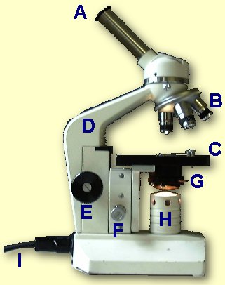
This is a common compound microscope. Label its parts from A ...
Compound Microscope - Diagram (Parts labelled), Principle and Uses A compound microscope basically consists of optical and structural components. Within these two systems, there are multiple components within them and they are: Image : Labeled Diagram of compound microscope parts See: Labeled Diagram showing differences between compound and simple microscope parts Structural Components

Compound Microscope Parts, Functions, and Labeled Diagram ...
Diagram of a Compound Microscope - Biology Discussion 1. It is noted first that which objective lens is in use on the microscope. 2. Stage micrometer is positioned in such a way that it is in the field of view. 3. The eyepiece is rotated so that the two scales, the eyepiece or ocular scale and the stage micrometer scale, are parallel. 4.
compound microscope with label and functions - Clip Art Library
quizlet.com › 522507578 › micro-module-1-flash-cardsMicro Module 1 Flashcards | Quizlet Study with Quizlet and memorize flashcards containing terms like Move the terms into the correct empty boxes to complete the concept map., Drag the images and/or statements to their corresponding class to test your understanding of the main types of microbes., Drag the images or descriptions to their corresponding class to test your understanding of the cellular organization and relative size ...

National Model 132-CLED Compound Microscope
en.wikipedia.org › wiki › ChloroplastChloroplast - Wikipedia A chloroplast / ˈ k l ɔːr ə ˌ p l æ s t,-p l ɑː s t / is a type of membrane-bound organelle known as a plastid that conducts photosynthesis mostly in plant and algal cells.The photosynthetic pigment chlorophyll captures the energy from sunlight, converts it, and stores it in the energy-storage molecules ATP and NADPH while freeing oxygen from water in the cells.
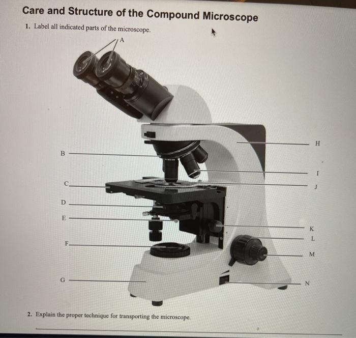
Solved Care and Structure of the Compound Microscope 1 ...
Compound Microscope Labeled Diagram | Quizlet Contains the ocular lens Body tube A hollow cylinder that holds the eyepiece. Arm Part that supports the microscope. Stage Supports the slide or specimen Coarse adjustment Knob sed to focus when using the low power objective lenses Fine Adjustment Knob Used to focus the image on high power to view image in more detail. Revolving nose piece
Parts of a microscope with functions and labeled diagram
Compound Microscope - Types, Parts, Diagram, Functions and Uses Image 2: The eyepiece/ocular lens of a compound microscope. Picture Source: slideplayer.com Eyepiece/ocular lens - It is the part of the microscope that is looked through at the top. It comes with a magnification ranging between 5x and 30x. Image 3: The head connects the eyepiece to the objective lens. Picture Source: microscope.com
Solved Label the image of a compound light microscope using ...
Compound Microscope- Definition, Labeled Diagram, Principle, Parts, Uses In order to ascertain the total magnification when viewing an image with a compound light microscope, take the power of the objective lens which is at 4x, 10x or 40x and multiply it by the power of the eyepiece which is typically 10x. Therefore, a 10x eyepiece used with a 40X objective lens will produce a magnification of 400X.

Compound Microscope Parts – Labeled Diagram and their ...
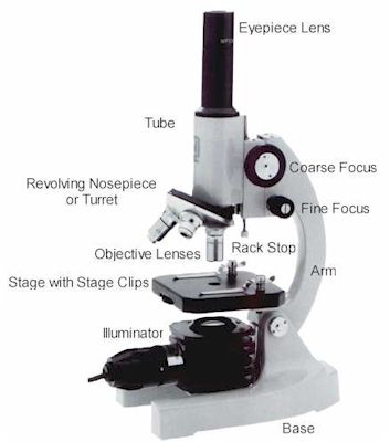
Microscope Parts and Functions Here are the important compound microscope parts... Eyepiece: The lens the viewer looks through to see the specimen. The eyepiece usually contains a 10X or 15X power lens. Diopter Adjustment: Useful as a means to change focus on one eyepiece so as to correct for any difference in vision between your two eyes.

Label The Parts Of A Compound Microscope Teaching Resources | TPT
› 2022/10/12 › 23400986Microsoft takes the gloves off as it battles Sony for its ... Oct 12, 2022 · Microsoft pleaded for its deal on the day of the Phase 2 decision last month, but now the gloves are well and truly off. Microsoft describes the CMA’s concerns as “misplaced” and says that ...

Compound microscope - their parts and function - Microscopy4kids
A Study of the Microscope and its Functions With a Labeled Diagram ... These labeled microscope diagrams and the functions of its various parts, attempt to simplify the microscope for you. However, as the saying goes, 'practice makes perfect', here is a blank compound microscope diagram and blank electron microscope diagram to label. Download the diagrams and practice labeling the different parts of these ...

How to draw compound of Microscope easily - step by step
› NATIONAL-GEOGRAPHIC-Dual-StudentNational Geographic Dual LED Student Microscope Aug 07, 2017 · Vanstarry Beginners Microscope Kit 40X-1000X for Kids & Students, Dual LED Lights and Cordless Capability, Illumination Lab Compound Monocular Microscopes with Optical Glass Lenses & 12 Slides 4.4 out of 5 stars 116
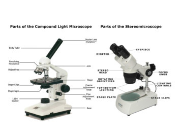
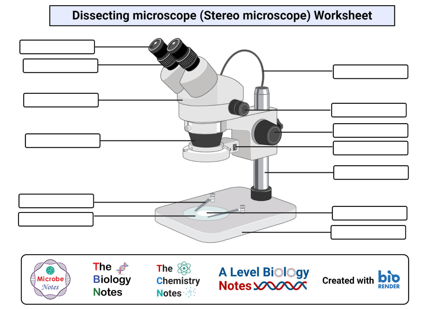
Stereo & Compound Microscope Print Out by CalmAndConfidence | TpT

AmScope - 40X-1000X Cordless LED Student Biological Compound Microscope + Slide Preparation Kit + World of The Microscope Book - M100C-LED-SP14-WM

Compound Microscope Labeled Diagram | Quizlet

Compound Microscope Parts, Diagram Definition, Application ...

A Study of the Microscope and its Functions With a Labeled ...
Parts of a Microscope with Their Functions – Microbe Online
Compound Microscope Parts, Functions, and Labeled Diagram ...
Parts Of A Microscope - Parts Of A Compound Microscope, HD ...
Microscope

List: Parts of a Microscope and their Function | Pathwooded
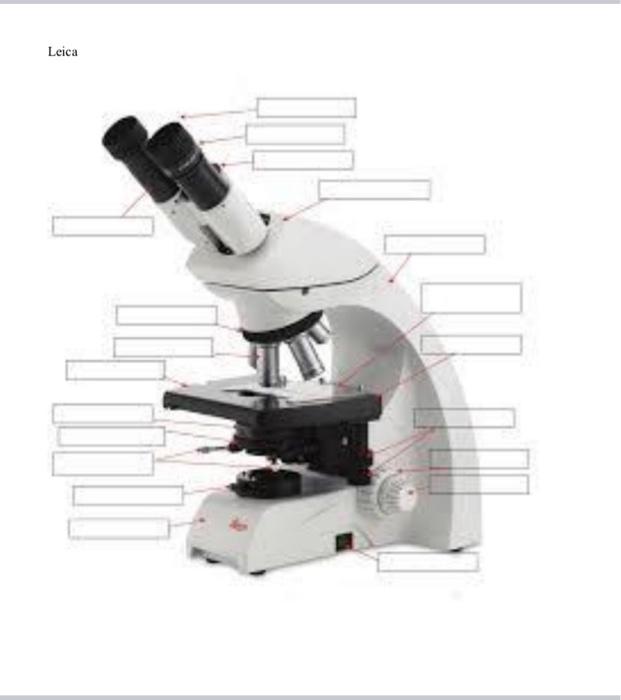
Solved Nikon Parts of the compound microscope Write the ...
Label a microscope - Teaching resources

Simple Microscope - Diagram (Parts labelled), Principle ...

Compound Microscope: Know Definition,working, diagram, properties

Care and Structure of the Compound Microscope 1.jpg - Care ...
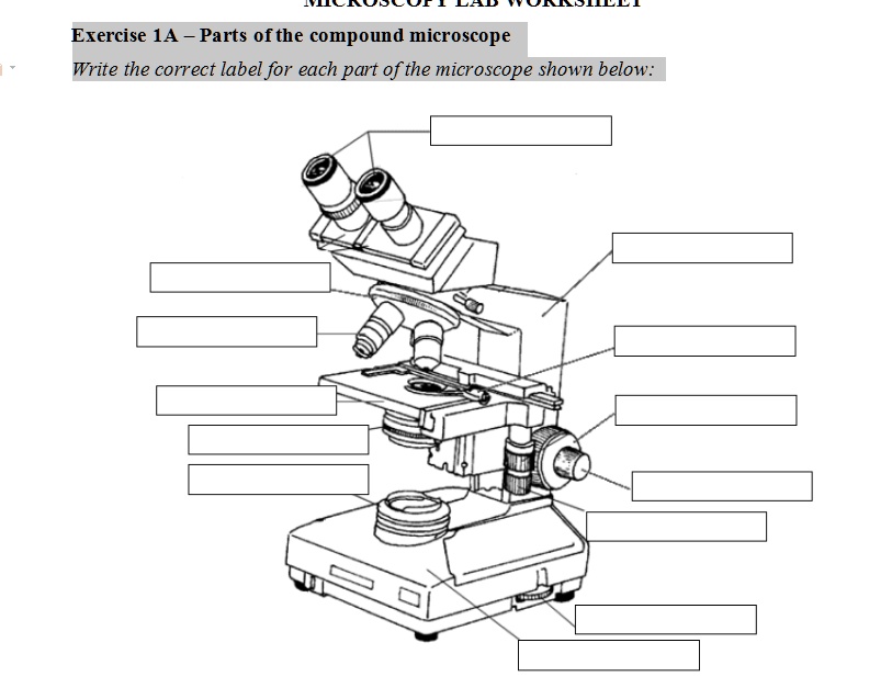
SOLVED: Exercise 1A Parts ofthe compound microscope Write the ...

Compound Microscope- Definition, Labeled Diagram, Principle ...

Living Environment Course

This is a common compound microscope. What the labelling D ...
What is Compound Microscope? - Diagram, Function, Advantages
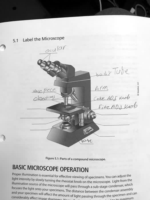
SOLVED: ' 5.1 Label the Microscope Ocu lar bad Tuhe Arm Lake ...

The Compound Microscope parts & how ... | Microscope parts ...

National 131-RLED-MS Compound Microscope with Mechanical Stage
Post a Comment for "44 picture of compound microscope with labels"